
Fluorescence for Automatic Seafloor Classification
Note – While the article below describes an application of fluorescence for automatic image classification in an underwater application, the concepts can apply to any use of fluorescence imaging. In addition to increasing contrast of a fluorescing subject against its background, fluorescence response can at times aid in distinguishing elements of an image from each other.
As you can tell from the many pictures in the Galleries, fluorescence can really make things stand out. You can spot tiny fluorescing critters, and you can often recognize what kind of organism something is based on its characteristic color or pattern. If it’s easy to do this by eye, there is also the potential to do this with computerized image processing techniques.
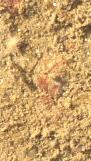
A number of years ago we collaborated with Robert Miller, who was then a graduate student at University of Massachusetts, Boston. He had deployed a set of granite settlement plates at a depth of about 10 meters near Nahant, Massachusetts. The surfaces were exposed to a range of conditions (shading, protection from grazers, etc.) to study what controlled the populations that developed. Robert took white-light photos and NIGHTSEA’s Charlie Mazel took fluorescence photos of the same area. As you can see in the photos below, there is a lot more to see with fluorescence. I wrote a classification routine to classify the features in the photographs into a few categories.
The first set of three images below are the white light, fluorescence, and computer-classified images for one of the settlement plates. The following three sets are blow-ups of portions of this image. The red fluorescence comes from algae that contain only chlorophyll, while the orange fluorescence comes from red algae that also contain phycoerythrin (see the article on sources of underwater fluorescence). The bright green may come from fluorescing subjects or reflected excitation light, while the source of the bright yellow is uncertain.


We have also applied this approach to a coral reef environment. In this case the fluorescence image was not made with a conventional camera, but with a sophisticated system called the Fluorescence Imaging Laser Line Scan (FILLS). FILLS swept a blue laser beam over the bottom and collected information in 4 channels – reflected blue light, green fluorescence (primarily from corals), orange fluorescence (from red algae, cyanobacteria, and some corals), and red fluorescence (from chlorophyll). The three fluorescence channels were combined into a false-color RGB image – red fluorescence to the Red channel, green to Green, and orange to Blue. We then used a computer classification routine to assign image pixels to one of 6 categories or ‘unknown’.
The three images below show the reflected blue light channel, the false-color RGB fluorescence image of the same scene, and the result of the computer separation into classes. The image covers a reef area of about 8 x 8 meters (26 x 26 feet).


Researchers are beginning to look at fluorescence photography as a way to improve classification of seafloor images. A group from Scripps Institution of Oceanography has developed a sophisticated image processing techniques for automatically annotating conventional white-light coral reef images (see CoralNet). A recent online article about their work ends with a discussion of adding fluorescence to improve accuracy.
Dr. Dave Zawada of the US Geological Survey and NIGHTSEA founder Charlie Mazel recently published a paper further exploring the potential to use fluorescence for automated classification. We looked at how the characteristic fluorescence emissions of different types of reef organisms might be recorded at different distances from the sea floor, and how many spectral bands might be needed to perform the classification.