
Hirox Fluorescence Images
The NIGHTSEA Model SFA adapter system adds a versatile fluorescence capability to the Hirox digital microscope. It can be used in a wide variety of applications, as illustrated by the examples below. Scroll down, or jump to any section of interest. Visit our Hirox product page for more information on the system and to request a quotation.
- Electronic component failure analysis
- Circuit boards – conformal coating and other fluorescence
- Anti-counterfeit – driver’s license
- Concrete thin section
- Epoxy on a small motor shaft
- Gel defect in a nylon granule
- Naturally fluorescent mineral
- Bone fragment in sand
Electronic component failure analysis
Examining electronic components that was embedded in epoxy, cross-sectioned, polished, and highlighted with a fluorescent epoxy-like dye. While the manufacturer of the fluorescent dye recommended excitation with ultraviolet light, our Royal Blue light head was used for these images, and was found to be superior to ultraviolet. You can read more about this kind of inspection application in the article on this web site.

Fluorescent penetrant highlighting cracks in integrated circuit, 80x, white light (top) and fluorescence under Royal Blue excitation
Circuit boards – conformal coating and other fluorescence
Conformal coating on a circuit board, with damage. Images made with Ultraviolet excitation (left) and white light.

Conformal coating on circuit board, 20x, fluorescence under UV excitation and white light light
Circuit board, white light and fluorescence under Royal Blue excitation


-

- 20x

-

- 60x

Anti-counterfeit – driver’s license
Images of the fluorescent markings on a driver’s license, made visible with ultraviolet light. These hidden markings on US licenses are generally quite different from state to state.
-

- 20x

-

- 60x

-

- 120x

-

- 160x

-

- 20x

-

- 20x

-

- 20x

-

- 20x

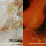
Concrete thin section
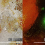
The images below were made from a 20μm concrete thin section with the NIGHTSEA Royal Blue excitation. As with the electronic components above, the manufacturer of the fluorescent dye recommended excitation with ultraviolet light. Our Royal Blue light head was used for these images and was found to be superior to ultraviolet. You can read more about this kind of inspection application in the article on this web site.
-
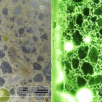
- Concrete thin section, 20x, white light and fluorescence under Royal Blue excitation

-
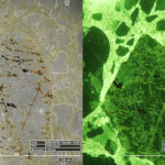
- Concrete thin section, 100x, white light and fluorescence under Royal Blue excitation

Thin section courtesy of Department of Mineralogy and Geochemistry, Institute of Geoscience and Geography, University Halle-Wittenberg, Germany.
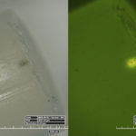
Epoxy on a motor shaft
Small motor shaft with epoxy where it does not belong. Imaged with white light (left) and ultraviolet excitation, 20x magnification.
-
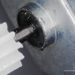
- Epoxy on motor shaft, 20x, white light

-
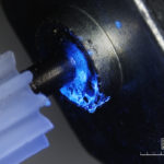
- Epoxy on motor shaft, 20x, UV excitation

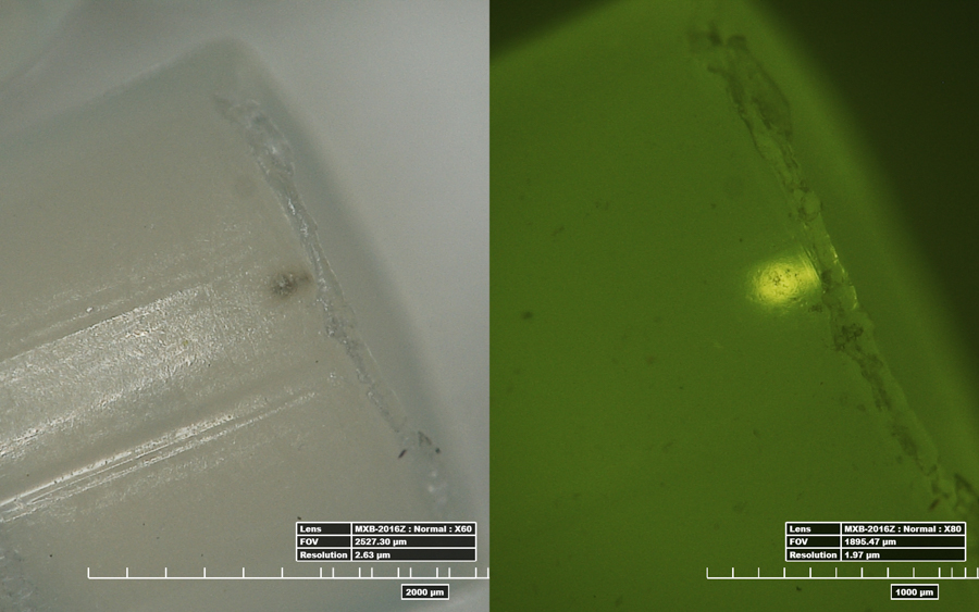
Gel defect in a nylon granule
Nylon 6,6 granules can manifest a process defect called ‘gel’ and if there is too much of this in a production batch it can compromise downstream production. The gel shows up as a brighter fluorescent area within the fluorescing granule. You can read more about this in our application article. Images made with Royal Blue excitation.
-
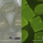
- Nylon granules with gel defect, 20x, white light and fluorescence under Royal Blue excitation

-
- Nylon granules with gel defect, 60x, white light and fluorescence under Royal Blue excitation
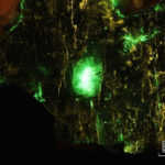
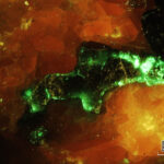
Naturally fluorescent mineral
This mineral sample, collected at the Sterling Hill Mine in Ogdensburg, New Jersey, contains willemite (green fluorescence), calcite (red fluorescence) and franklinite (black – no fluorescence).
-
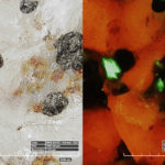
- Fluorescent mineral, 20x, white light and fluorescence under Royal Blue excitation

-
- Fluorescent mineral, 60x, white light and fluorescence under Royal Blue excitation

-
- Fluorescent mineral, 100x, white light and fluorescence under Royal Blue excitation

-
- 20x, blue light

-
- 20x, blue light

Bone fragment
Small bone fragment in sand.

Bone in sand, 20x, fluorescence under UV excitation (top) and white light