
Keyence VHX Fluorescence Images
The NIGHTSEA Model SFA adapter system adds a versatile fluorescence capability to the Keyence VHX Digital Microscope series, and the combination is being used in an ever-expanding variety of applications. The adapter works with many of the available Keyence lenses, including the VH-Z00, -Z20, -Z50, -Z100, -Z500, -ZST, and the VHX-7101 (FI). Visit our Keyence product page for more information.
The images below are a sampling of those we have made during our own development and testing or in collaboration with customers. Scroll down, or jump to any section of interest.
- Electronic component failure analysis
- Circuit board conformal coating
- Concrete thin section
- Epoxy on a small motor shaft
- Gel defect in a nylon granule
- Maize and periwinkle
- Mosquito midguts
- Fluorescent-dosed plastic particles
- Naturally fluorescent mineral
- Ammonite fossil
- Wax verification
Electronic component failure analysis
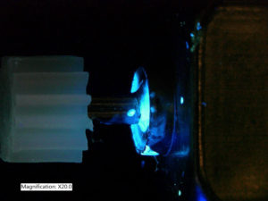
Examining electronic components that were embedded in epoxy, cross-sectioned, polished, and highlighted with a fluorescent epoxy-like dye. Images were made with the Keyence VH-ZST lens. While the manufacturer of the fluorescent dye recommended excitation with ultraviolet light, our Royal Blue light head was used for these images, and was found to be superior to ultraviolet. You can read more about this kind of inspection application in the article on this web site.
-

- Component 1

-

- Component 2

-

- 50x

-

- 100x

-

- 300x

-

- 500x

-

- 1000x

-

- 2000x

-

- 30x

-

- 50x

-

- 100x

-

- 200x

-

- 300x

-

- 500x

-

- 1000x

-

- 1000x

-

- 2000x

-

- 2000x

Circuit board conformal coating
Conformal coating on a circuit board, with damage. Images made with Keyence VH-ZST lens. The upper row was made with Ultraviolet light, the ‘traditional’ wavelength range for this application. While this works well with the excitation directed from the side, the ultraviolet wavelengths do not work well with the ZST lens when the light head is positioned over the fiber optic input port. The lower row shows the same areas under excitation with our Violet light head. While the fluorescence is marginally less bright at lower magnifications, this source works better through the Keyence lens optics, producing the brighter area you can see in the 200x image.
Ultraviolet excitation
-

- 20x

-

- 30x

-

- 50x

-

- 100x

-

- 200x

Violet excitation
-

- 20x

-

- 30x

-

- 50x

-

- 100x

-

- 200x

Concrete thin section
The images below were made from a 20μm concrete thin section with the Keyence VH-Z100 lens, NIGHTSEA Royal Blue excitation. As with the electronic components above, the manufacturer of the fluorescent dye recommended excitation with ultraviolet light. Our Royal Blue light head was used for these images and was found to be superior to ultraviolet. You can read more about this kind of inspection application in the article on this web site.
-

- 100x

-

- 200x

-

- 500x

-

- 700x

Thin section courtesy of Department of Mineralogy and Geochemistry, Institute of Geoscience and Geography, University Halle-Wittenberg, Germany.
Epoxy on a motor shaft
Small motor shaft with epoxy where it does not belong. Imaged with Keyence VH-ZST lens, 20x magnification.
-
- Epoxy on motor shaft, 20x

Gel defect in a nylon granule
Nylon 6,6 granules can manifest a process defect called ‘gel’. If there is too much gel in a production batch it can compromise downstream production. You can read more about this in our application article.
Images made with Keyence VH-ZST lens, Royal Blue excitation. At 100x and above the images utilized the Keyence automated focus stacking capability.
-

- 30x

-

- 50x

-

- 100x, with focus stacking

-

- 200x

-

- 300x

-

- 500x

Maize and periwinkle
The images below were shared by Dr. Amy Clore, Professor of Biology at New College of Florida. Both images were made with a Keyence VHX-7000 and the VH-Z100 lens. That lens allows direct coupling of the NIGHTSEA excitation light source into the lens optics, which can provide enhanced performance with biological samples such as these.
The image on the left shows a fresh slice of maize (Zea mays L.) endosperm stained with Sytox Green. This reagent binds preferentially to nuclei and to a lesser degree to the cell periphery.
This image on the right shows a periwinkle (Catharanthus roseus L.) bud that has been dissected so that all of its floral organ types are visible. At this stage these organs contain chlorophyll, which emits the distinctive red autofluorescence seen here.
-
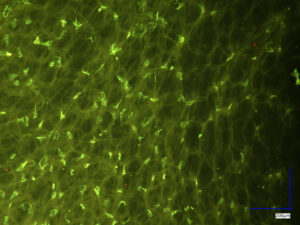
- Maize (Zea mays L.) stained with Sytox Green

-
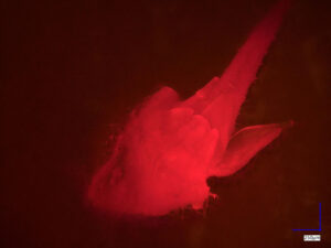
- Natural chlorophyll fluorescence of periwinkle (Catharanthus roseus L.) bud

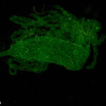
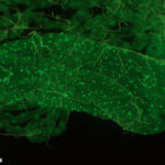
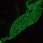
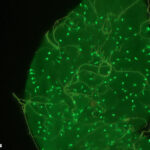
Mosquito midguts
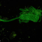
This is a nice example of imaging of a biological subject with the Keyence microscope. The images are of the midguts of female Aedes aegypti mosquitoes, mounted on a microscope slide. The samples were immunostained with a rabbit antiserum for neuropeptide F (1/500) and goat anti-rabbit antibody conjugated to Alexa 488. The fluorescent green cells are isolated peptide producing endocrine cells. The samples were generously provided by the Department of Entomology, University of Georgia, Athens, GA.
The images were made with a Keyence VHX-7000 microscope with the FI (VHX-7101) multi-objective head, using the NIGHTSEA Royal Blue excitation/emission set.
-
- Sample 1, 50x

-
- Sample 1, 80x

-
- Sample 1, 150x

-
- Sample 2, 100x

-
- Sample 3, 300x, focus stacked

Fluorescent-dosed plastic particles
Fluorescent-dosed plastic particles, 38 – 45 microns. Image made with ultraviolet excitation. Image courtesy of an explosives detection company.

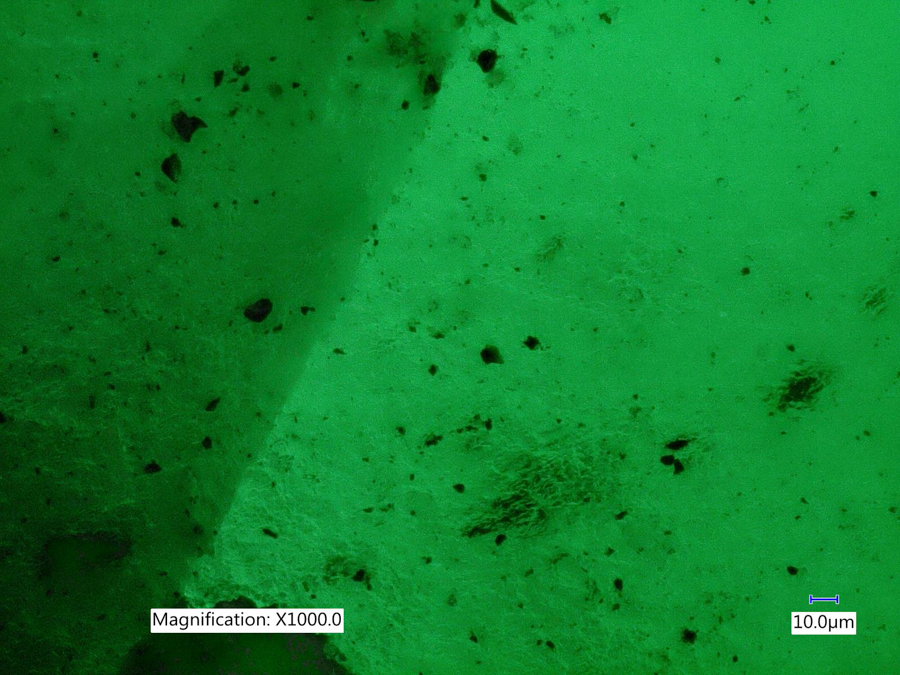
Naturally fluorescent mineral
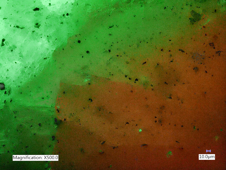
This mineral sample, collected at the Sterling Hill Mine in Ogdensburg, New Jersey, contains willemite (green fluorescence), calcite (red fluorescence) and franklinite (black – no fluorescence).
-
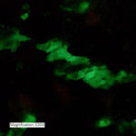
- Willemite, 20x

-
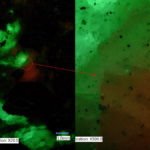
- Willemite/calcite boundary, composite 20x and 500x

-
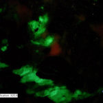
- Willemite/calcite boundary, 20x

-
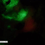
- Willemite/calcite boundary, 50x

-
- Willemite/calcite boundary, 500x
-
- Willemite, composite 20x and 1000x

-
- Fluorescent mineral – Willemite, 2000x

-
- Willemite, 1000x
Ammonite fossil
Sectioned and polished fossil of an ammonite, an early (and now extinct) cephalopod, related to modern-day nautilus, squid, cuttlefish, and octopus.
-

- White light

-

- Fluorescence

Ammonite fossil fluorescing under the Keyence microscope, VH-ZST lens, Royal Blue excitation.
-

- 20x

-

- 50x

-

- 100x

-

- 200x

-

- 500x

Wax verification
Verifying proper wax (torque modifier) application on parts from a subcontractor under UV excitation.
-

- Wax verification under UV excitation

-

- Wax verification under UV excitation
