Fluorescence images of juvenile corals
Posted On: Friday, November 7, 2014
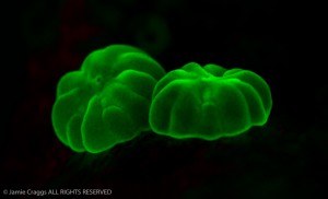
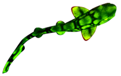
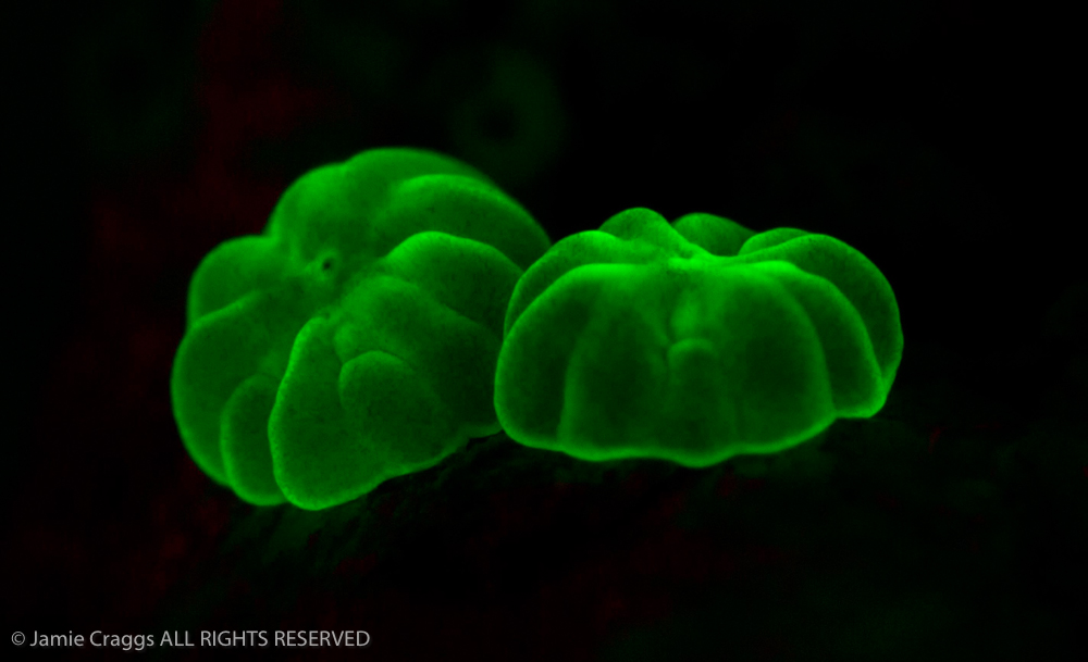
These beautiful close-up images of tiny (~1mm) coral polyps were contributed by Jamie Craggs, aquarium curator at the Horniman Museum & Gardens in London. You would normally expect images like this from a stereo microscope, but Jamie took these handheld with a Canon 5D with a Canon MP-E 65mm macro lens set for 5x, two Inon Z240 strobes with NIGHTSEA EX-INON excitation filters, and a yellow barrier filter in front of the camera lens. Thanks Jamie!
Fluorescence is an invaluable tool for coral recruitment research in the field, the aquarium, and the laboratory.
-
- Newly settled coral polyps
-
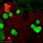
- Newly settled coral polyps, fluorescence (c) Jamie Craggs, Horniman Museum & Gardens

I wonder why the Aquarium of Fluorescent Corals in Noumea, New Caledonia, is never mentioned when fluorescent corals are written about.
Hi Phalbe. The work of Dr. Rene Catala at the Noumea Aquarium is certainly recognized. Visit the Articles page (accessed in the Resources section on the menu bar), scroll down to the Marine Science or Reef Aquarium or Sport Diving section and go to the article HIstory of Underwater Fluorescence Observation and Photography (https://nightsea.com/articles/underwater-fluorescence-history/). The 4th paragraph is all about Dr. Catala.
That doesn’t mean there couldn’t be more here about Dr. Catala and the Aquarium itself. If you would like to write something up I would be happy to post it.