Pre-Screening Samples for Fluorescence
The NIGHTSEA Model SFA Stereo Microscope Fluorescence Adapter can turn your routine laboratory stereo microscope into a valuable tool for pre-screening your sample preparations for fluorescence before moving on to higher resolution systems.
[You can also download this application note in pdf format]
The Challenge
High resolution imaging of biological samples is heavily based on fluorescence techniques. Confocal, 2-photon, and high resolution compound fluorescence microscopes are almost always a limited resource. They are often located only in imaging core facilities and accessible on a scheduled, pay-per-use basis.
The processes for introducing fluorophores to specimens are not always successful. Staining, introduction of GFP-bearing plasmids to cells, immunohistochemistry – all are fallible. It is not unusual to spend time searching for fluorescence on a high end system when there is not even any there to be found.
The Practical Solution
The NIGHTSEA SFA enables fluorescence pre-screening of specimens on a standard stereo microscope. The detail that you see is not important – the simple presence or absence and general location of fluorescence lets you know whether it is worth taking your specimen to the imaging core. Between the direct expense of the use fee and the time wasted to look at a non-fluorescent specimen it will not take many saved trips for the NIGHTSEA system to more than pay for itself.
One researcher’s work requires staining rabbit psoas muscle fibers with Alexa Fluor 488 Phalloidin. There was some frustration with samples that did not take up the stain. After acquiring the SFA she wrote:
“The NIGHTSEA fluorescence setup is a great way to quickly check whether the stain was successful before we try to image the muscle fiber at a higher magnification on the confocal.”
(Click image for larger view)

Rabbit psoas muscle fibers stained with Alexa Fluor 488 Phalloidin, in white light and fluorescence. Images made using NIGHTSEA’s white LED (left) and the Royal Blue excitation/emission light+filter set. Samples courtesy of Dr. Beth Brainerd and Natividad Chen, Brown University.
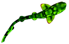
Another researcher uses zebrafish as a system to look at the way different toxicants (pharmaceuticals, pesticides, food additives, etc.) alter brain development. He writes:
“Before using NIGHTSEA to screen my samples, I would have to select samples to mount, go to the confocal and then hope that some of my samples were actually fluorescent. Now that I use NIGHTSEA to prescreen my samples I save both time and money by making sure the only samples I image are fluorescent.”
(Click image for larger view)

Confocal image of brain of transgenic zebrafish (Dania rerio). Kaede protein – green is unconverted, red is photoconverted. Image courtesy of Robert Thorn, Creton Lab, Brown University.
Download the application note in pdf format.